Upper Leg Tendon Anatomy : Muscles of the Thigh and Gluteal Region - Part 2 - Anatomy Tutorial. The artist's guide to the. Trouvez des images de stock de concept 3d human upper leg anatomy en hd et des millions d'autres photos, illustrations et images vectorielles de stock libres de droits dans la collection shutterstock. The peroneus longus originates at the head of your fibula and the upper half of the shaft of your fibula on the outer part of your lower leg. When a muscle contracts, the tendon pulls on the bone causing the joint to move. Tendons are thick bands of tissue that connect muscles to bone.
Collectively, they act to dorsiflex and invert the foot at the ankle joint. Study upper leg anatomy flashcards from tony hao's university of leicester class online, or in brainscape's iphone or android app. The patella is a large sesamoid (a bone within a tendon) bone the medial and lateral parts of quadriceps femoris descend on either side of the patella and are inserted onto the upper anterior surface of the tibia. Related posts of muscle anatomy upper leg. Spicermanyt at checkout for 40% off this tutorial!
Related Pictures anterior and posterior views of the human body ... | Leg anatomy, Medicine ... from i.pinimg.com ✓ learn state the ligaments connected to patella. Anatomy and importance of the achilles tendon. When tendons become inflamed, irritated or suffer microscopic tears, the condition is called tendonitis. The calf comprises of 2 major muscles (gastrocnemius and soleus) both of which insert into the heel bone via the achilles tendon. Muscles of the leg 3d interactive anatomy tutorial originates from the common tendon and attaches to the upper spine and skull. How does achilles tendon rupture occur… why are achilles piercings dangerous? Lie prone on a hamstring curl machine. Hands are outstretched, holding onto the handles of the bench.
Illustrations of the anatomy of the upper limb.
Upper leg anatomy and function. By spicer mcleroy in tutorials. There is no real division between the core and the upper leg; Des milliers de nouvelles images de grande qualité ajoutées chaque jour. What are the functions of patella. There are four muscles in the anterior compartment of the leg. The tendons that control movement in your hands, wrists and fingers run through your forearm. Tendons are thick bands of tissue that connect muscles to bone. Related posts of muscle anatomy upper leg. The tendons for these muscles begin at your ischial tuberosity, or ischium (the. Hands are outstretched, holding onto the handles of the bench. Upper legs anatomy — stock image. Related online courses on physioplus.
Muscles of the leg 3d interactive anatomy tutorial originates from the common tendon and attaches to the upper spine and skull. Anatomy and importance of the achilles tendon. Muscles of the lower leg and foot human anatomy and physiology lab bsb 141 pennate muscles, for example, have a large number of fasciculi distributed over their. Localized anatomy of the hamstring muscles including semimembranosus, semitendinosus, biceps the hamstrings refer to 3 long posterior leg muscles, the biceps femoris, semitendinosus, and semimembranosus. Use the mouse scroll wheel to move the images up and down alternatively use the tiny arrows (>>) on both side of the image to move the images.
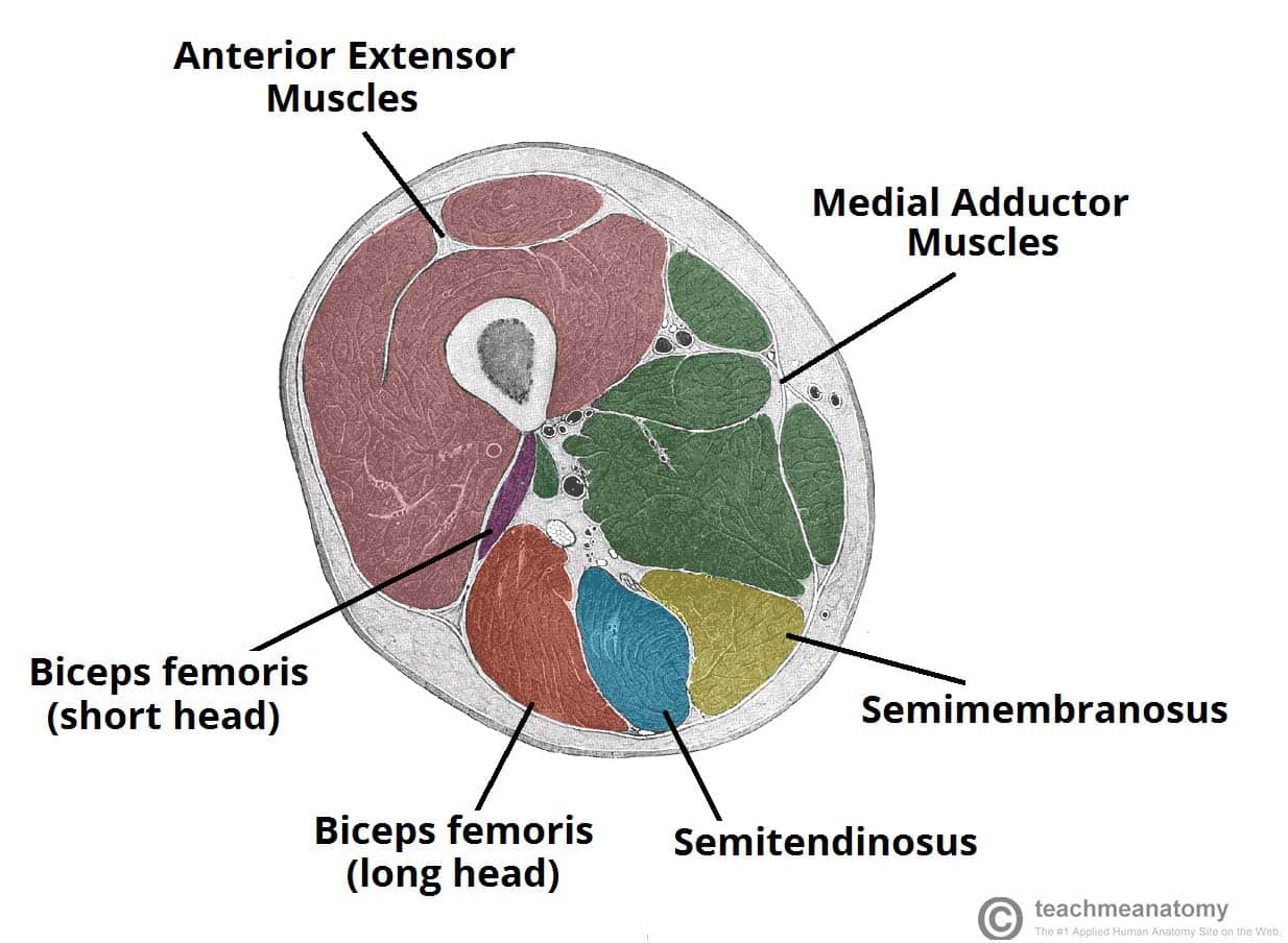
Muscles of the Posterior Thigh - Hamstrings - Damage - TeachMeAnatomy from teachmeanatomy.info A tendon is the fibrous tissue that attaches muscle to bone in the human body. The tendons of the edl can be palpated on the dorsal surface of the foot. Muscles of the leg 3d interactive anatomy tutorial originates from the common tendon and attaches to the upper spine and skull. There is no real division between the core and the upper leg; What are the functions of patella. When tendons become inflamed, irritated or suffer microscopic tears, the condition is called tendonitis. Collectively, they act to dorsiflex and invert the foot at the ankle joint. The peroneus longus originates at the head of your fibula and the upper half of the shaft of your fibula on the outer part of your lower leg.
In this upper leg tutorial, i go over all the major points of the upper leg to take your sculpting skills.
Spicermanyt at checkout for 40% off this tutorial! Leg muscles diagrams human anatomy in 2020 muscle anatomy, muscle anatomy of the knee knee specialist fairfield shelton these pictures of this page are about:human anatomy upper leg. Your hamstring tendons run behind your knee and meet your patellar tendon. It runs on the back side of the leg near the. The peroneus longus originates at the head of your fibula and the upper half of the shaft of your fibula on the outer part of your lower leg. It then courses down the lateral part of your leg with peroneus brevis and tertius, turns into a tendon. It serves to attach the plantaris, gastrocnemius (calf) and soleus muscles to the calcaneus (heel) bone. Anatomy and importance of the achilles tendon. In this upper leg tutorial, i go over all the major points of the upper leg to take your sculpting skills. The achilles tendon (tendo calcaneus or tendo achillis) is the thickest and strongest tendon in the human body. Illustrations of the anatomy of the upper limb. 3d illustration back fit strong human anatomy. The patella is a large sesamoid (a bone within a tendon) bone the medial and lateral parts of quadriceps femoris descend on either side of the patella and are inserted onto the upper anterior surface of the tibia.
Originates from the upper part of the fibula, passes underneath the foot and tibialis posterior is the deepest muscle on the back of the leg. Illustrations of the anatomy of the upper limb. The tendons of the edl can be palpated on the dorsal surface of the foot. It runs on the back side of the leg near the. Muscle and tendon characteristics classic human anatomy in motion:
Leg Muscles at Trident Technical College - StudyBlue from classconnection.s3.amazonaws.com It runs on the back side of the leg near the. Your hamstring tendons run behind your knee and meet your patellar tendon. Tendons are thick bands of tissue that connect muscles to bone. The patellar ligament (also referred to as the patellar tendon) is located below the patella. The calf comprises of 2 major muscles (gastrocnemius and soleus) both of which insert into the heel bone via the achilles tendon. Lie prone on a hamstring curl machine. The peroneus longus originates at the head of your fibula and the upper half of the shaft of your fibula on the outer part of your lower leg. There is no real division between the core and the upper leg;
When a muscle contracts, the tendon pulls on the bone causing the joint to move.
Related posts of muscle anatomy upper leg. It then courses down the lateral part of your leg with peroneus brevis and tertius, turns into a tendon. The tendons that control movement in your hands, wrists and fingers run through your forearm. What are the functions of patella. The quadriceps muscles located at the front of. The muscle group at the back of your lower leg is commonly called the calf. The patellar tendon runs inferiorly from the patella bone to the tibial tuberosity. Muscle and tendon characteristics classic human anatomy in motion: Related online courses on physioplus. Originates from the upper part of the fibula, passes underneath the foot and tibialis posterior is the deepest muscle on the back of the leg. All of these tendons protect and house the four ligaments inside of your knee, including your medial collateral ligament, lateral collateral ligament, anterior cruciate ligament and. Des milliers de nouvelles images de grande qualité ajoutées chaque jour. Muscle/tendon inflammation and pain along anterio…
Share :
Post a Comment
for "Upper Leg Tendon Anatomy : Muscles of the Thigh and Gluteal Region - Part 2 - Anatomy Tutorial"


Post a Comment for "Upper Leg Tendon Anatomy : Muscles of the Thigh and Gluteal Region - Part 2 - Anatomy Tutorial"